Visualizing a protein decision complex in actin filament length control

Seen at the Gelles Lab Little Engine Shop blog this week, commentary on a new paper in Nature Communicationspublished in collaboration with the Goode Lab and researchers from New England Biolabs.

“Single-molecule visualization of a formin-capping protein ‘decision complex’ at the actin filament barbed end”

[ensemblevideo contentid=Z33TDbsofEW7ofOXsSti8w autoplay=true showcaptions=true]

Regulation of actin filament length is a central process by which eukaryotic cells control the shape, architecture, and dynamics of their actin networks. This regulation plays a fundamental role in cell motility, morphogenesis, and a host of processes specific to particular cell types. This paper by recently graduated [Biophysics and Structural Biology] Ph.D. student Jeffrey Bombardier and collaborators resolves the long-standing mystery of how formins and capping protein work in concert and antagonistically to control actin filament length. Bombardier used the CoSMoS multi-wavelength single-molecule fluorescence microscopy technique to to discover and characterize a novel tripartite complex formed by a formin, capping protein, and the actin filament barbed end. Quantitative analysis of the kinetic mechanism showed that this complex is the essential intermediate and decision point in converting a growing formin-bound filament into a static capping protein-bound filament, and the reverse. Interestingly, the authors show that “mDia1 displaced from the barbed end by CP can randomly slide along the filament and later return to the barbed end to re-form the complex.” The results define the essential features of the molecular mechanism of filament length regulation by formin and capping protein; this mechanism predicts several new ways by which cells are likely to couple upstream regulatory inputs to filament length control.

Single-molecule visualization of a formin-capping protein ‘decision complex’ at the actin filament barbed end
Jeffrey P. Bombardier, Julian A. Eskin, Richa Jaiswal, Ivan R. Corrêa, Jr., Ming-Qun Xu, Bruce L. Goode, and Jeff Gelles
Nature Communications  6:8707 (2015)

The capping protein expression plasmid described in this article is available from Addgene.

Readers interested in this subject should also see a related article by Shekhar et al published simultaneously in the same journal.  We are grateful to the authors of that article for coordinating submission so that the two articles were published together.

Easy Come, Easy Go

Whereas the diffusion of water molecules in the bulk liquid depends entirely on breaking hydrogen bonds, the diffusion of proton defects (i.e., an excess proton in acid or a proton deficit in base) is expedited by proton hopping across hydrogen bonds.  The details of this process are well understood in acid, and the process in base was believed to occur in analogous fashion. However, theoretical studies of hydroxide have given highly divergent predictions of solvation structures and diffusion rates, depending on the chosen recipe for such simulations: some predicted the traditionally expected solvation structures and some predicted the experimentally observed diffusion trends, but none do both. Now Seyit Kale, a graduate student in Prof. Judith Herzfeld’s group, has studied proton defects using the group’s recently developed LEWIS force field.[1] The LEWIS simulations obtain the correct relative diffusion rates with hydroxide solvation structures that are analogous to those of hydronium,[2] thereby supporting the traditional picture of the “proton hole”. The authors also catch and characterize proton transfer events, identifying similar “special pairs”[3] as the intermediates in both cases (see figure).

[1]       S. Kale, J. Herzfeld, J. Chem. Phys. 2012, 136, 084109.
[2]       S. Kale, J. Herzfeld, Angew. Chem. Int. Edit. 2012 in press. DOI: 10.1002/anie.201203568.
[3]       O. Markovitch, H. Chen, S. Izvekov, F. Paesani, G. A. Voth, N. Agmon, J. Phys. Chem. B. 2008, 112, 9456-9466.


Molecular mechanisms of noisy transcription

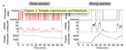
In a recently published paper “Effect of promoter architecture on the cell-to-cell variability in gene expression” in PLoS Computational Biology, Alvaro Sanchez and co-workers investigated how the architecture of a model promoter region (characterized by number of transcription factor binding sites, the binding affinity and spacing on the DNA) affects the way in which individual cells respond to environmental stimuli. In particular, they examine, using stochastic chemical kinetics, how the intrinsic randomness in the binding and unbinding of transcription factors to their binding sites generates cell-to-cell differences in transcript and protein levels within a population. The analysis uses a combination of computational modeling and analytical mathematical methods. Sanchez, a recent Ph.D. graduate in Biophysics and Structural Biology performed this work with Jané Kondev (Physics), and in collaboration with Rob Phillips, Hernan Garcia and Daniel Jones (Caltech).

While previous population-average models explained well how promoter architecture affects the average response of a population of cells to changes in the concentration of transcription factors, the question of how the response of individual cells is determined by promoter region sequence remains generally unsolved and limited to simplified coarse-grain models. By way of an example, the authors of this study investigated the effect of cooperative binding between transcription factors in the level of variability in the transcriptional response to increasing concentrations of those factors. It is well known that cooperativity in gene regulation increases the sensitivity of the response of the promoter to changes in the intracellular concentration of transcription factors, leading to a switch-like response. By examining this architecture, Sanchez and co-workers found that cooperativity is also a source of large intrinsic cell-to-cell variability in gene expression: larger sensitivity comes accompanied with larger variability (even if all cells contain the exact same amount of repressor).

This investigation continues a collaboration between theorists at Brandeis and experimentalists at Caltech, which aims to connect the biochemical, molecular understanding of transcriptional regulation coming from in vitro biochemical experiments (which are also being done in the Gelles lab at Brandeis) with the phenotypic behavior of individual cells as determined by gene expression measurements in single live cells. Many of the predictions of this computational study are currently being tested in Rob Phillips’ lab.