First Rosbash-Abovich Award Recipients Announced

Michael Rosbash, the Peter Gruber Endowed Chair in Neuroscience and Professor of Biology and his wife, Nadja Abovich, established the Rosbash-Abovich Award as a way to inspire and acknowledge excellence in research by post-doctoral fellows and graduate students in the Brandeis life sciences. The Rosbash-Abovich award will be awarded annually.

The award honors the most outstanding papers published the previous year that have been authored by a Brandeis postdoctoral fellow and a Brandeis PhD student. In addition to the honor being selected, each winner is presented with a monetary award.

Future winners will present their talks at upcoming Volen Scientific Retreats, but due to COVID restrictions, the 2020 winners will be presenting their talks during the Molecular Genetics Journal Club meetings.

Most outstanding paper by a post-doctoral fellow

Michael O'Donnell
Michael O’Donnell, PhD

The 2020 winner for the most outstanding post-doctoral paper is Michael O’Donnell for the publication titled “A neurotransmitter produced by gut bacteria modulates host sensory behavior“. O’Donnell, is a former postdoc in the Piali Sengupta Lab. Sengupta said

Mike is a remarkable scientist and mentor. He single-handedly and independently established a new research direction in my lab. He also served as an informal mentor to many graduate students and has continued to do so even after he left my lab. I greatly appreciated our long discussions and arguments, and he is very much missed.

Sengupta also noted that O’Donnell was chosen to receive this award

on the basis of the creativity and novelty of his work that was published in Nature. The committee was particularly interested in nominating a researcher who was a driving force behind the work and Mike certainly fulfilled this criteria.

O’Donnell is now an assistant professor at Yale and recently formed the O’Donnell lab. He presented his talk to the Molecular Genetics Journal Club on December 2, 2020. He spoke about his work on neuromodulators produced by different bacteria.

Most outstanding paper by a PhD student

James Haber & Gonen Memisoglu
Professor James Haber & Gonen Memisoglu, PhD

The recipient of the 2020 award for the most outstanding PhD student paper is Gonen Memisoglu for the publication “Mec1 ATR Autophosphorylation and Ddc2 ATRIP Phosphorylation Regulates DNA Damage Checkpoint Signaling.“ She was a PhD student in James Haber’s lab. She received her PhD in 2018 and is currently a postdoctoral fellow at the University of Chicago. She will be presenting her talk at the Molecular Genetics Journal Club on February 2, 2021.

When asked about his former PhD student, Haber said

I was delighted to learn that Gonen was the recipient of the Rosbash/Abovich award for the best publication by a graduate student last year; but I had to ask “which paper” because Gonen made two important discoveries last year about the way cells respond to DNA damage. Gonen helped develop a highly efficient way to edit the yeast genome and to create dozens of very precise mutations in the Mec1 gene that is the master regulator of the DNA damage response.  When there is a chromosome break, the Mec1 protein phosphorylates a number of proteins that creates a cascade of signaling to prevent cells from progressing through mitosis until damage is repaired. Gonen discovered that the extinction of the this signal depended on Mec1’s autophosphorylation of one specific target and that changing that specific amino acid to one that could not be phosphorylated was enough to cause cells to remain arrested. She also identified several alterations of the Ddc2 protein that associates with Mec1 that were also critical for its normal activation.

During her time in my lab Gonen was a super hard-working and exceptionally insightful grad student, but also incredibly generous with her time, helping others in the lab

The Benefits of Middle Age

Almost all our cells harbor a sensory organelle called the primary cilium, homologous to the better known flagella found in protists. Some of these cilia can beat and allow the cell to move (eg. in sperm), or move fluid (eg. cerebrospinal fluid) around them. However, other specialized cilia such as those found in photoreceptor cells and in our olfactory neurons function solely as sensory organelles, providing the primary site for signal reception and transduction. The vast majority of our somatic cells display a short and simple rod-like cilium that plays crucial roles during development and in adulthood. For instance, during development, they are essential for transducing critical secreted developmental signals such as Sonic hedgehog that is required for the elaboration of cell types in almost every tissue (eg. in brain, bones, muscles, skin). In adults, cilia are required for normal functioning of our kidneys, and primary cilia in hypothalamic neurons have been shown to regulate hunger and satiety.

Given their importance, it is not surprising that defects in cilia structure and function lead to a whole host of diseases ranging from severe developmental disorders and embryonic lethality to hydrocephalus (fluid accumulation in the brain), infertility, obesity, blindness, and polycystic kidney among others. Often these diseases manifest early in development resulting in prenatal death or severe disability, but milder ciliary dysfunction leads to disease phenotypes later in life.

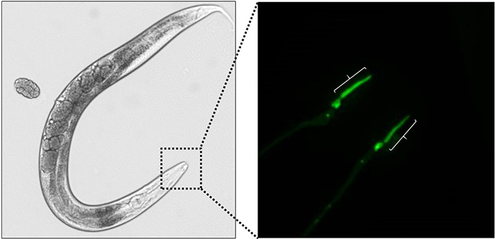
Much is now known about how cilia are formed and how they function during development. However, surprisingly, how aging affects cilia, and possibly the severity of cilia-related diseases, is not well studied. A new study by postdocs Astrid Cornils and Ashish Maurya, and graduate student Lauren Tereshko from Piali Sengupta’s laboratory, and collaborators at University College Dublin and University of Iowa, begins to address this question using the microscopic roundworm C. elegans (pictured below). These worms display cilia on a set of sensory neurons; these cilia are built by mechanisms that are similar to those in other animals including in humans. Worms have a life span of about 2-3 weeks, thereby making the study of how aging affects cilia function quite feasible.

benefits-midage

They find that cilia structure is somewhat altered in extreme old age in control animals. However, unexpectedly, when they looked at animals carrying mutations that lead to human ciliary diseases, the severely defective cilia seen in larvae and young adults displayed a partial but significant recovery during middle-age, a period associated with declining reproductive function. They went on to show that the heat-shock response and the ubiquitin-proteasome system, two major pathways required for alleviating protein misfolding stress in aging and neurodegenerative diseases, are essential for this age-dependent cilia recovery in mutant animals. This restoration of cilia function is transient; cilia structure becomes defective again in extreme old age. These results suggest that increased function of protein quality control mechanisms during middle age can transiently suppress the effects of some mutations in cilia genes, and raise the possibility that these findings may help guide the design of therapeutic strategies to target specific ciliary diseases. Some things can improve with aging!

Form meets function

Professor of Biology Piali Sengupta gave the 2015 Stetten Lecture at NIGMS on Oct 21 on Form Meets Function: Structurally Diverse Cilia and Their Roles in Sensory SignalingThe “cilia squad” in the Sengupta Lab has been working for some years now to examine cilia formation in sensory neurons in the nematode C. elegans, and the relationship between the structure and nerve cell function. You can watch Piali’s lecture online.


Deep inside a worm’s nose

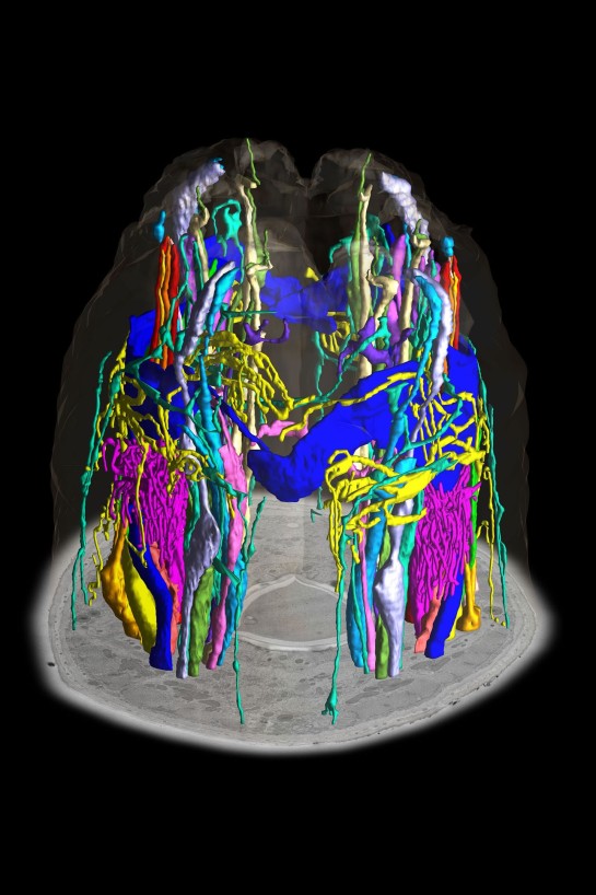
In a new paper in eLIFE, a team spearheaded by Brandeis postdocs David Doroquez and Cristina Berciu provide a strikingly detailed look at key structures called cilia on neurons involved in sensory perception in the nematode C. elegans. Primary cilia are the antenna-like structures onsensory neurons that gather information about the animal’s environment, such as chemicals, temperature, humidity, and touch. The genetic tools available to manipulate individual, identifiable neurons in C. elegans make worms an excellent model organism to study the assembly and function of cilia. This study requires a description of the structure of the cilia and their immediate surrounding glial support cells, and this new paper, a collaboration of the Sengupta and Nicastro labs, provides high-resolution 3D models showing how diverse and specialized these structures are.

worm-01-2
A bouquet of sensory antennae. The 3D ultrastructure of all sensory cilia
and other neuronal projections in the head of the soil roundworm C.
elegans have been reconstructed using serial section transmission electron
microscopy. Shown are 3D isosurface-rendering models emerging from a
transmission electron microscopic cross-section of the worm.

The key techniques in this study were serial section transmission electron microscopy and electron tomography, with structures well-preserved by high-pressure freezing and freeze-substitution. With these techniques, the authors achieved the first high-resolution 3D reconstructions of 50/60 cilia from C. elegans. They describe several previously uncharacterized features — for example, there are distinct types of branching patterns – in one, the two cilia originate from independent basal bodies (as previously seen in Chlamydomonas). In the second, the cilia branch after the basal transition zone, the ciliary gatekeeper region. In the latter case, this basically means that whatever is needed for the cilia to branch has to be transported through the transition zone, suggest there might be novel mechanisms of ciliary protein trafficking. In a third pattern, the branching occurs proximally before the transition zone, and represent therefore dendritic microvilli, rather than ciliary branching. The study also showed different organizations  of microtubules in different cilia types and vesicles in regions of the cilia which have never been seen before, again pointing to new mechanisms of protein transport. They also describe new cilia-glial interactions, which might suggest that cilia and glia talk to each other.

For more about these structures (with lots of pretty pictures and movies), see:

More science

We’ve all been busy this spring writing grants and teaching courses and doing research and graduating(!), so lots of publications snuck by that we didn’t comment on. Here’s a few I think that might be interesting to our readers.

  • From Chris Miller‘s lab, bacterial antiporters do act as “virtual proton efflux pumps”:
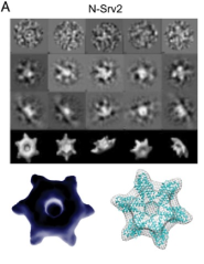
  • nsrv2Are ninja stars responsible for controlling actin disassembly? Seems like the Goode lab might think so.
    • Chaudhry F, Breitsprecher D, Little K, Sharov G, Sokolova O, Goode BL. Srv2/cyclase-associated protein forms hexameric shurikens that directly catalyze actin filament severing by cofilin. Mol Biol Cell. 2013;24(1):31-41.
  • What do you get from statistical mechanics of self-propelled particles? The Hagan and Baskaran groups team up to find out.
  • From John Lisman and Ole Jensen (PhD ’98), thoughts about what the theta and gamma rhythms in the brain encode
  • From Mike Marr‘s lab, studeies using genome-wide nascent sequencing to understand how transcrption bursting is controlled in eukaryotic cells
  • From the Lau and Sengupta labs, RNAi pathways contribute to long term plasticity in worms that have gone through the Dauer stage
    • Hall SE, Chirn GW, Lau NC, Sengupta P. RNAi pathways contribute to developmental history-dependent phenotypic plasticity in C. elegans. RNA. 2013;19(3):306-19.
  • Can nanofibers selectively disrupt cancer cell types? Early results from Bing Xu‘s group.
    • Kuang Y, Xu B. Disruption of the Dynamics of Microtubules and Selective Inhibition of Glioblastoma Cells by Nanofibers of Small Hydrophobic Molecules. Angew Chem Int Ed Engl. 2013.

How does a hard-wired simple circuit generate multiple behaviors?

In a paper appearing in last week’s issue of Neuron, members of the Sengupta Lab and their collaborators from the Bargmann Lab describe how a fixed neural circuit produces multiple behaviors in a context-dependent manner.  The study was led by former Brandeis post-doctoral fellow Kyuhyung Kim in the Sengupta Lab (currently Assistant Professor at DGIST, Korea) and Rockefeller student Heeun Jang in the Bargmann Lab. Also involved in the study were current Brandeis MCB students Scott Neal and Danna Zeiger, and Dongshin Kim, the head of the Brandeis Microfluidics Facility.

For this study the researchers used the nematode Caenorhabditis elegans. The nervous system of C. elegans consists of only 302 neurons (in the adult hermaphrodite) whose anatomical connectivities are well-mapped. Despite its relatively small nervous system, C. elegans exhibits a wide range of behaviors in response to environmental stimuli. For instance, C. elegans exhibits varied responses to pheromones – small chemical substances used for intra-specific communication. Some pheromones are repulsive to adult hermaphrodite C. elegans but neutral to male C. elegans. However, reducing the function of the neuropeptide Y-like receptor NPR-1 results in hermaphrodites now exhibiting neutral pheromone responses and males becoming strongly attracted. The researchers asked how the sex and neuromodulatory state of the animal allows it to interpret the pheromone stimulus differently to generate distinct behavioral responses.

To answer this question, the researchers used behavioral assays, genetic manipulations of neuronal output, and in vivo measurements of pheromone-induced neuronal activity (using genetically encoded calcium sensors and customized microfluidics devices designed by the Brandeis Microfluidics Facility). They found that flexible output of a neuronal ‘hub-and-spoke’ circuit motif was responsible for generating these distinct pheromone responses under different conditions.

In this circuit, pheromone-sensing neurons ASK and ADL are connected to the central RMG motor/interneuron by gap junctions (see Figure). Jang et al. showed that in hermaphrodites with high levels of NPR-1 activity, the ADL sensory neurons respond strongly to a specific pheromone component and drive avoidance behavior via their chemical synapses to command interneurons for locomotion. However, sexual dimorphism in the circuit results in males having reduced ADL pheromone responses.  Moreover, Jang et al. showed that ADL synaptic output in males is further decreased via RMG and ASK-mediated antagonism (see Figure). As a result, males are indifferent to this pheromone.

The next issue the authors addressed is the role of NPR-1 activity in regulating pheromone responses. The Bargmann Lab had previously shown that high NPR-1 activity inhibits RMG, and under these conditions, pheromone responses of the ASK sensory neurons are low. Conversely, when NPR-1 activity is reduced or absent, ASK pheromone responses are enhanced. Jang et al. found that in the absence of NPR-1 activity, ADL chemical synaptic output in response to pheromones is antagonized by the RMG-ASK gap junction circuit. In other words, avoidance mediated by ADL chemical synaptic output is balanced by attraction mediated by the RMG-ASK gap junction circuit, resulting in hermaphrodites being neither attracted to nor avoiding this pheromone. In males with reduced NPR-1 activity the same effects are observed, however, since the ADL pheromone response is already lower in males, the RMG-ASK attraction-mediating arm “wins” resulting in attraction to pheromones.  The authors refer to these as overlapping ‘push-pull’ circuits in analogy with electronic circuits.

These results begin to explain how a small fixed circuit can generate a remarkable range of behaviors via alteration of sensory response properties as well as choice of specific synaptic output pathway as a function of neuromodulatory state and sex. The general theme of a circuit functioning differently under different neuromodulatory conditions has been extensively studied in the Marder Lab in the crustacean nervous system, and is an important principle to be kept in mind when interpreting functionality from structurally described connectomes.

Jang H(*), Kim K(*), Neal SJ, Macosko E, Kim D, Butcher RA, Zeiger DM, Bargmann CI, Sengupta P. Neuromodulatory State and Sex Specify Alternative Behaviors through Antagonistic Synaptic Pathways in C. elegans. Neuron. 2012;75(4):585-92.

How many neurons does it take to stay cool?

The worm (nematode) C. elegans is a nice model system for studying neuroscience, combining

  • genetic tools that allow genes to be turned on or off, often on a per cell basis, in the whole organism,
  • tools like laser or genetic ablation that allow individual, identified cells to be selectively eliminated,
  • robust behaviors that can be readily measured, and
  • a well defined nervous system consisting of 302 neurons, each of which can be identified, and whoseanatomical connectivity has been established.

In a paper appearing this month in Journal of Neuroscience, Molecular and Cell Biology grad student Matthew Beverly, undergrad Sriram Anbil, and Professor of Biology Piali Sengupta examined the contribution of sensory neurons to controlling thermotaxis in C. elegans. Worms develop a memory of the temperature at which they have been cultivated, and display a robust behavior in which worms placed on a temperature gradient at temperatures higher than their cultivation temperature will crawl back towards colder temperature (negative thermotaxis – see movie). The behavior depends on TAX-4, a channel protein expressed in a subset of the sensory neurons. In this study, the Brandeis researchers asked the question “how many and which of the sensory neurons are required for the worms to perform negative thermotaxis, and are the required sensory neurons the same regardless of the temperature range examined?” (or, in my paraphrase, “how many and which neurons does it take to stay cool?”)

Worm head, showing expression of the calcim indicator GCaMP in ASI and AWA neurons (used in calcium imaging experiments)

As it turns out, the answer is complicated (and readers are encouraged to read the paper). The researchers found that in addition to the previously known thermosensory neurons AFD and AWC, the ASI neurons previously known to be involved in chemosensation play a significant role in regulating negative thermotaxis. Interestingly, the circuits used seem to be degenerate; under one condition, for example, a particular combination of AFD, AWC or ASI is necessary to generate the behavior, although at other conditions, a different combination is required to generate the same behavior.. And only a couple of degrees Celsius makes a difference — the circuit required for negative thermotaxis on a gradient centered at 8oC above the cultivation temperature is different from a gradient centered at 6oC above.

These and other results taken together suggest that even in the worm, a complex circuit has evolved to control crawling behaviors to cope with temperature changes, and that having degeneracy in the underlying circuits may be a common feature that ensures that behaviors crucial to survival are maintained in a variety of environmental conditions..

Beverly M, Anbil S, Sengupta P. Degeneracy and Neuromodulation among Thermosensory Neurons Contribute to Robust Thermosensory Behaviors in Caenorhabditis elegans. J Neurosci. 2011;31(32):11718-27.

Time for Worms in Circadian Biology

Almost every organism on earth, from archae to humans, exhibits circadian rhythms – periodic cycles of behavior or gene expression that repeat approximately every 24 hours. These rhythms are generated by a circadian clock – an internal time-keeping mechanism – which can be entrained and synchronized by environmental cues such as temperature or light/dark cycles. This clock may provide organisms with an adaptive advantage throughout their life, and disruption of the function of this clock can lead to severe behavioral and metabolic disorders in humans.

For more than two decades researchers have wondered whether the tiny soil-dwelling nematode worm Caenorhabditis elegans, one of the foremost model organisms, contains a circadian clock. Circadian rhythmic behaviors described previously in C. elegans are variable and hard to quantify, and no genes were known to exhibit gene expression oscillations with 24 hr cycles as shown in many other animals.

Now, in a recent study published in the open-access journal PLoS Biology, several students and postdoctoral fellows in the labs of Piali Sengupta and Michael Rosbash joined forces and took on the challenge to identify C. elegans genes under clock control.

Light and temperature cycles both drive and entrain 24 hr oscillations in gene expression in C. elegans.

They showed that indeed C. elegans contains genes whose expression cycles in a circadian manner. They found that light and temperature cycles appear to regulate different sets of genes (see above), indicating that these stimuli may entrain two distinct clocks. Moreover, the underlying clock mechanisms may not be dependent on oscillations of known clock genes. “These findings were surprising to us since Drosophila only has a single conserved clock running in multiple cells and tissues” says Alexander van der Linden – lead author and former postdoctoral fellow in the Sengupta Lab.

C. elegans has a wealth of genetic and behavioral tools. The next critical step will be to identify the mechanisms underlying the C. elegans circadian clock(s). These investigations may also provide information of how the clock evolved since nematodes and humans split about 600-1200 million years ago.

Alexander M. van der Linden is now an Assistant Professor at the University of Nevada, Reno. The work was conducted in the labs of Profs. Michael Rosbash, a member of the Howard Hughes Medical Institute and Piali Sengupta in the Department of Biology. Other authors who contributed to this work include Molecular and Cell Biology graduate students Matthew Beverly, Joseph Rodriquez and Sara Wasserman (now a postdoctoral fellow at UCLA), and Sebastian Kadener, a former postdoctoral fellow who is now an Assistant Professor at the Silberman Institute of Life Science, The Hebrew University of Jerusalem, Israel.

Brandeis professor and postdoc alumni win Searle Scholars grants

One Brandeis professor and two of our former postdocs are among the 15 recipients of the competitive Searle Scholars awards for 2010. Nelson Lau, an assistant professor in the Biology Dept., won for his work on “Germline Genome Regulation by Piwi Proteins and Small RNAs”. Katie Henzler-Wildman is a former postdoc from the Kern lab in Biochemistry who now has a lab at Washington Univ. in St. Louis. Katie received a grant to study “The Role of Protein Dynamics in Multidrug Resistance Transport Activity”. David Biron, an alumnus of the Sengupta Lab who is now an assistant professor at U. Chicago, received a grant to study “Understanding Lethargus: The Sleep-Like Behavior of the Nematode C. Elegans“.

Congratulations to all the winners!

Research quickies

Some of our recent publications (descriptions are mine, not the authors’)

Lau: Finding new insect viruses by sequencing small RNAs (siRNA and piRNA)

Katz Lab: Taste affects smell

Sengupta Lab: Stress early in life causes epigenetic changes in worms