Locus coeruleus catecholamines link neuroticism and vulnerability to tau pathology in aging

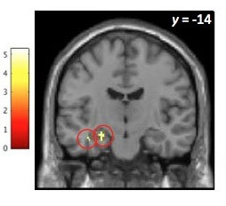
More than 6 million people in the U.S. are living with Alzheimer’s disease in 2022. The prevalence of this neurodegenerative disease has prompted scientists to study the factors that may increase someone’s risk for developing Alzheimer’s disease. Higher neuroticism is a well-known dementia risk factor, which is associated with disordered stress responses. The locus coeruleus, a small catecholamine-producing nucleus in the brainstem, is activated during stressful experiences. The locus coeruleus is a centerpiece of developing models of the pathophysiology of Alzheimer’s disease as it is the first brain region to develop abnormal tau protein, a hallmark feature of the disease. Chronic activation of stress pathways involving the locus coeruleus and amygdala may promote tau spread, even in cognitively normal older adults. This leads to the question of whether high-neuroticism individuals show non-optimal affective function, altered locus coeruleus neurotransmitter function, and greater tau accumulation.  Researchers in the Neurochemistry and Cognition Lab, led by Dr. Anne Berry set out to answer this question.LC blog post figurePhD candidate Jourdan Parent examined relationships among personality traits, locus coeruleus catecholamine neurotransmitter function, and tau burden using positron emission tomography imaging in cognitively normal older adults. She found that lower locus coeruleus catecholamine function was associated with higher neuroticism, more depressive symptoms, and higher tau burden in the amygdala, a brain region implicated in stress and emotional responses. Exploratory analyses revealed similar associations with low trait conscientiousness, a personality trait that is also considered a risk factor for dementia. Path analyses revealed that high neuroticism and low conscientiousness were linked to greater amygdala tau burden through their mutual association with low locus coeruleus catecholamine function. Together, these findings reveal locus coeruleus catecholamine function is a promising marker of affective health and pathology burden in aging, and that this may be a candidate neurobiological mechanism for the effect of personality on increased vulnerability to dementia.


Locus coeruleus catecholamines link neuroticism and vulnerability to tau pathology in aging. Jourdan H.Parent, Claire J.Ciampa, Theresa M. Harrison, Jenna N. Adams, Kailin Zhuang, Matthew J.Betts, Anne Maass, Joseph R. Winer, William J. Jagust, Anne S. BerryNeuroImage, 30 September 2022, 119658.


Blanchette and Scalera et al., discover new insights into an intercellular communication method in neurons

Fruit fly neuron (magenta) with extracellular vesicle cargoes (green). Cargoes are packaged inside the neuron and, then released outside of the neuron in extracellular vesicles.

Research scientist Cassie Blanchette and Neuroscience Ph.D. student Amy Scalera, working in the Rodal lab, discovered a new mechanism of regulation of extracellular vesicles (EVs). EVs are small, membrane-bound compartments that can transfer cargoes such as DNA and proteins between cells for communication. EVs are important for normal cell-cell signaling, but they are also hijacked in neurodegenerative disease to spread toxic disease proteins to other cells. Therefore, it is crucial to understand how and where EVs are formed. Blanchette and Scalera discovered a novel method of regulation of EVs specifically at the synapses (the region of the neuron that contacts adjacent cells), using the fruit fly nervous system as an experimental model.

EVs are derived from endosomes, a network of intracellular sorting compartments that cells use to separate cargoes into different ‘packages’ with distinct inter and intracellular destinations. Blanchette and Scalera found a surprising function for the proteins that regulate endocytosis, a process in which the cell membrane buds inward, thus forming a compartment to bring cargoes to endosomes. The authors found that mutants lacking endocytic proteins lose the local pool of EV cargoes that are available for release from synapses, and instead send these cargoes for disposal elsewhere in the neuron. They hypothesized that the normal function of endocytosis  is akin to a plane circling in a holding pattern at an airport – while it waits for its time to land, it is better for the passengers to circle (between the cell membrane and endosomes), nearby their destination (release in EVs), rather than being sent to an entirely different city (a different region of the neuron). They also found that disrupting this holding pattern had consequences for the physiological functions of EV cargoes; in endocytic mutants, loss of Synaptotagmin-4, an EV cargo important for neuronal adaptability, was associated with failure of the neuron to grow in response to firing. Endocytic mutants also caused synaptic depletion of the Alzheimer’s disease associated EV cargo Amyloid Precursor Protein (APP), thus suppressing its toxicity and increasing the survival of APP-expressing flies. These discoveries raise the possibility that proteins regulating EV traffic may be targets for neurodegenerative disease therapies.

Turrigiano lab uncovers sources of neuronal heterogeneity

High activity neurons have greater instrinsic excitability and response to local inputs, but no difference in total input type or amount

Mammalian cortex has long been one of the most widely studied systems in neuroscience, dating back to the pioneering work of Santiago Ramon y Cajal in the late 19th century. The cortex is much larger in primates than other mammals, and is thought to be responsible for the advanced cognitive abilities of humans. Today, models of cortical connections and computations form the basis for some of the most powerful deep learning paradigms. However, despite this success, there is still much that is unknown about how cortex functions. One feature of cortex that has recently been discovered is that neurons that appear to be similar to each other can have very different baseline activity levels: some neurons are 100x more active than their neighbors. We don’t know how neurons that are otherwise highly similar in shape and genetic makeup can maintain such different activity levels, or if the neurons with high and low activity levels have different functions in the brain. These neurons are otherwise so similar to each other that it is difficult to tell them apart without recording their activity directly, and current techniques for recording the activity of many neurons simultaneously in live animals do not allow us to later re-identify them for further study.

In a paper recently published in Neuron, the Turrigiano lab, led by postdoctoral researcher Nick Trojanowski, reported a new approach for permanently labeling high and low activity neurons in live animals, and then determining what makes them different. To do this they used a fluorescent protein called CaMPARI2 that changes from green to red as activity increases, but only when exposed to UV light. By shining UV light into the brain, they caused neurons with high activity to turn red, while neurons with low activity remained green. This procedure allowed them to run a series of tests on high and low activity neurons to identify differences between them. They found that high activity neurons would intrinsically generate more activity than low activity neurons when presented with the same stimulus. These high activity neurons also receive more excitatory input specifically from nearby neurons of the same type. Surprisingly, however, they found that the total amount of excitatory and inhibitory input that high and low activity neurons received from other neurons was not a major factor in determining their activity levels. Together, these results tell us that the differences in activity between neurons are due to intrinsic differences, as well as their pattern of connectivity to their nearby partners. This has deep implications for how the networks that underlie cortical computations are built and maintained.

With these tools in hand, it is now possible to further explore the differences between high and low activity neurons. Do these neurons serve different functions? Are the baseline activity levels specified from birth? How do these activity levels affect the mechanisms of plasticity that are responsible for learning and memory? The recently published results represent just the tip of the iceberg of information that can be learned with this new technique, in the mammalian cortex as well as other brain regions.

Leading Science: Magnifying the Mind

Brandeis Magnify the Mind

Written by Zosia Busé, B.A. ’20

Joshua Trachtenberg, graduated from Brandeis in 1990 and is a leader in studying the living brain in action using advanced imaging technology. After establishing his research laboratory at UCLA, he founded a company – Neurolabware – in order to build the sophisticated custom research microscopes that are needed to perform groundbreaking work in understanding how the brain develops and how diseases and injuries interrupt its normal functioning. His company is created by scientists and for scientists, and is unique in creating high quality microscopes that are easy to use but also have the flexibility to be used in creative ways in innovative experiments, and in combination with a variety of other devices.

Brandeis University is now seeking to acquire one of these advanced microscopes that can observe fundamental processes inside the living brains of animals engaged in advanced behaviors. The resonant scanning two-photon microscope from Neurolabware allows researchers to understand and image large networks of neurons in order to visualize which cells and networks are involved with specific memories or how these processes go awry in disease. “This approach is unparalleled. There is no other technique around that could possibly touch this,” Trachtenberg says.

Previous two-photon technologies permitted only very slow imaging, allowing scientists to take a picture about every two seconds, but the resonant two-photon technology is a major breakthrough that allows scientists to take pictures at about 30 frames per second. This speed increase is a major game changer. Not only can one observe activity in the brain at a higher speed, but it is possible to take pictures at a speed that is faster than the movement artifacts that must be accounted for, such as breathing or heart beats. Because one can see the movement, it can be corrected, allowing high resolution functional imaging of structures as small as single synaptic spines in the living brain. Further, advances in laser technology and fluorescent labels now allow scientists to see deeper into the brain than ever before, compounding the recent advantages of increased speed.

Continue reading “Leading Science: Magnifying the Mind”

Cross-Cultural Differences in Brain Activity of Specific and General Recognition

Results from paper
Results revealed regions in the left fusiform (left circle) and left hippocampus (right circle) emerged when comparing activity for correct same versus correct similar responses across cultures.

A recent publication from Paige, Ksander, Johndro, & Gutchess (Cortex, 2017) of the Aging, Culture, and Cognition Lab at Brandeis University has shed light on how culture affects brain activation when encoding information into memory. Prior work has suggested that culture influences how people perceive the world, including how much perceptual detail (e.g., size, shape, color, etc.) is remembered. It may not be surprising that culture shapes customs or even social interactions, but evidence also suggests that it shapes cognition. Because encoding details into memory necessitates the engagement of additional cognitive resources, comparing across cultures on the specificity of memory offers a glimpse into which processes and types of information are considered important across cultural groups.

Participants who originated from America or East Asia studied photos of everyday items in a magnetic resonance imaging (MRI) scanner and 48 hours later completed a surprise recognition test. The test consisted of same (i.e., previously seen in the scanner), similar (i.e., same name, different features; for example, a coffee mug that is a different shape or color than what the participant saw at encoding), or new photos (i.e., items not previously seen in the scanner) and participants were instructed to respond “same,” “similar,” or “new.”

Unlike other studies, culture did not disproportionately influence behavioral memory performance for specific information. However, East Asians showed greater activation in the left fusiform and left hippocampus relative to Americans for specific (items correctly recognized as same) versus general memory (items correctly recognized as similar). Additional follow-up analyses confirmed this cultural pattern was not driven by differential familiarity with the items across cultures. One possible explanation for this finding is cultural differences in prioritization of high (e.g., fine details, local information) versus low spatial information (e.g., coarser, global information). In the present study, increased activation in the left medial temporal regions for East Asians may be reflective of additional processes needed to encode specific details into memory, reflecting the greater demands of local, high spatial frequency processing. Current work in the lab is addressing this possibility.

Past work has failed to consider how cross-cultural differences can occur at both the behavioral and neural level. The present findings remedy that, suggesting that culture should be considered an individual difference that influences memory specificity and its underlying neural processes.

Paige, L. E., Ksander, J. C., Johndro, H. A., & Gutchess, A. H. (2017). Cross-cultural differences in the neural correlates of specific and general recognition. Cortex91, 250-261.


The Amygdala, Fraud and Older Adults

Figure from Zebrowitz-Gutchess paper
Figure 1. Peak amygdala activation as a function of face trustworthiness for older adult participants. Error bars represent standard errors. COPE is the contrast of parameter estimates [high or medium, or low trustworthy faces minus baseline fixation] from which peak values were extracted at the subject-level using FSL featquery. * p < .05.
There is a widespread belief that older adults are more vulnerable to consumer fraud than younger adults. Behavioral evidence supporting this belief is mixed, although there is a reliable tendency for older adults to view faces as more trustworthy than do younger adults.  One study provided supporting neural evidence by demonstrating that older adults failed to show greater amygdala activation to low than high trustworthy faces, in contrast to considerable evidence that younger adults do show this effect. This result is consistent with the argument for greater vulnerability to fraud in older adults, since the amygdala responds to threatening stimuli. More generally, however, the amygdala responds to biologically salient stimuli, and many previous studies of younger adults have shown that this includes not only threatening, low trustworthy faces, but also high trustworthy faces. The Zebrowitz Face Perception Lab therefore included medium trustworthy faces in order to detect separate effects of high trustworthiness and low trustworthiness on amygdala activation in older adults, something that the one previous study of older adults did not do. Consistent with that study we found that older adults did not show stronger amygdala activation to low than high trustworthy faces.  However, they did show stronger amygdala activation to high than to medium trustworthy faces, with a similar trend for low vs medium, although that difference was not strong enough to be confident that it would replicate (See Figure 1).

The fact that older adults did not show greater amygdala activation to low than medium or high trustworthy faces is consistent with the suggestion that older adults may be more vulnerable to fraud. However, an important question is whether vigilant responding to untrustworthy-looking faces could actually protect one from fraud.  Arguing against this possibility is the finding that although younger adults have consistently shown greater amygdala activation to people who look untrustworthy, they do not show greater activation to those who actually cheat.  On the other hand, some evidence indicates that facial appearance does provide valid cues to threat. Face shape not only influenced younger adults’ trust of potential exploiters, but it also proved to be a valid indicator of economic exploitation.  Furthermore, this face shape cue influenced both younger and older adults’ accurate impressions of aggressiveness. To shed further light on neural mechanisms for any age differences in vulnerability to fraud that may exist requires investigating: 1) the sensitivity of neural responses to actual differences in trustworthiness in the domain of economic exploitation, and 2) whether any age differences in those neural responses are related to differential vulnerability to economic exploitation.

Zebrowitz, L.A., Ward, N., Boshyan, J., Gutchess, A., & Hadjikhani, N. (2017).  Older adults’ neural activation in the reward circuit is sensitive to face trustworthiness.  Cognitve, Affective, and Behavioral Neuroscience.


Communicating Memory Information Between the Hippocampus and Prefrontal Cortex

Jadhav paper full image

The brain has a remarkable capacity to record our daily experiences and recall this stored information to guide our behavior. For example, every time you decide to get a cup of coffee on campus, you immediately know where to go and then step toward your destination. The ability to successfully memorize paths and navigate in the environment is fundamental for animals searching for food (see Illustration), as well as for humans surviving in a complicated environment, especially when you don’t have your smartphone to rely on, but only your brain as the inner GPS! However, how does the brain learn and remember such plans that allow us to get from one place to another?

We know that a structure in brain called the hippocampus plays an important role in encoding and storing memories. The hippocampus is thought to replay remembered experiences during fast, ripple-like brain waves, termed sharp-wave ripples (SWRs), that occur during “down-time” for the brain, i.e., offline periods during sleep and during pauses in active behavior. It has been previously shown by Jadhav and colleagues that selectively disrupting these ripple oscillations using precisely-timed electrical impulses impairs the ability of animals to learn in spatial mazes, suggesting that this “mental replay” is important for navigation and memory (Jadhav et al., 2012, Science). Notably, mental replay is not isolated activity in the hippocampus, but works together with the prefrontal cortex (PFC), the executive center of brain involved in storing memories and making decisions (Jadhav et al., 2016, Neuron). However, exactly how such memory replay supports memory processing in waking and sleep states had remained elusive.

In a new article published in the Journal of Neuroscience (Tang et al., 2017), the Jadhav lab (the team included Neuroscience graduate students Wenbo Tang and Justin Shin) used high-density electrophysiology to record large numbers of neurons in both the hippocampus and prefrontal cortex in both sleep and awake states. They discovered that as rats learned a spatial memory task, the activity in the hippocampal-prefrontal network replayed recent experiences in a precise manner during SWRs that occurred when animals paused from actively exploring the maze. This structured mental replay related to ongoing spatial behavior is ideally suited for storing and retrieving memories to inform decisions. When animals were asleep after exploring the maze, the hippocampal-prefrontal replay, however, appeared “noisy” and mixed. This replay occurring during sleep periods can support the ability of the brain to consolidate memories, by selectively integrating related memories to build a coherent map for long-term storage (see Illustration). These findings show how memory information is communicated between the hippocampus and PFC during ripple oscillations, and indicate that mental replay during sleep and awake states serve distinct roles in memory. These studies collectively provide fundamental knowledge about the neural substrates of memories. They will thus provide important insights into memory deficits that are prevalent in many neurological disorders that involve the hippocampal-prefrontal network, such as Alzheimer’s disease and schizophrenia.

Hippocampal-Prefrontal Reactivation during Learning Is Stronger in Awake Compared with Sleep States. Wenbo Tang, Justin D. Shin, Loren M. Frank and Shantanu P. Jadhav. Journal of Neuroscience 6 December 2017, 37 (49) 11789-11805.


William T. Newsome to Receive 2015 Pepose Award on March 18

William NewsomeWilliam T. Newsome, a Stanford neuroscientist, will receive the 6th annual Jay Pepose ’75 Award in Vision Science on March 18. Newsome will deliver a lecture, “A New Look at Gating: Selective Integration of Sensory Signals through Network Dynamics,” on March 18 at 4:00 PM. The lecture will be held in Gerstenzang 121 and is open to the public.

Professor Newsome’s research at Stanford has helped scientists better understand the connection between visual perception and visually guided behavior. Newsome is the Harman Family Provostial Professor at the Stanford School of Medicine and is the Vincent V.C. Woo Director of the Stanford Neuroscience Institute.

The Pepose Award is funded by a $1 million endowment through a gift from Brandeis alumni Jay Pepose ’75, MA’75, P’08, P’17, and his wife,  Susan K. Feigenbaum ’74, P’08, P’17, through the Lifelong Vision Foundation. The endowment also supports graduate research fellowships in vision science.

Learn more

Shantanu Jadhav Wins Sloan Research Fellowship

Shantanu Jadhav

Shantanu Jadhav, assistant professor of Psychology and Neuroscience and one of our newest faculty members, has won the prestigious Sloan Research Fellowship from the Alfred P. Sloan Foundation.

Jadhav’s research focuses on how the hippocamus and the prefrontal cortex interact and communicate with each other.  This activity influences the brain’s ability to learn, remember and make decisions.

More information about Shantanu Jadhav’s research and the Sloan Research Fellowship can be found at Brandeis NOW.


SPROUT grant opportunity for 2015 announced

From the Brandeis Office of Technology Licensing:

The Brandeis Virtual Incubator invites members of the Brandeis Community (faculty, staff and students) to submit an application for the SPROUT Program. These Awards are intended to stimulate entrepreneurship on campus and help researchers launch their ideas and inventions from the lab to the marketplace.The SPROUT Program will provide pilot funding for innovative scientific projects within the Division of Science that require bench research, lab space, and/or lab equipment.

We will be awarding $50,000 to be shared among the most promising proposals.
Come get your questions answered at one of our upcoming information sessions.
Info Sessions: 
Thursday, February 26,  11:00 a.m.-12:00 p.m. (Volen, room 201)
Monday, March 2,  2:00 p.m.-3:00 p.m.   (Shapiro Science Center, 1st Floor Library, room 1-03)
 
Deadlines: Preliminary Proposals are due by Friday, March 6th
Please note, the introduction of the new SPARK Program geared towards innovative non-bench projects that have impact. An additional email will be sent detailing this program.
For more information on each program go to our website or contact the OTL program leaders,  Melissa Blackman for SPROUT and  Anu Ahuja  for SPARK.