Communicating Memory Information Between the Hippocampus and Prefrontal Cortex

Jadhav paper full image

The brain has a remarkable capacity to record our daily experiences and recall this stored information to guide our behavior. For example, every time you decide to get a cup of coffee on campus, you immediately know where to go and then step toward your destination. The ability to successfully memorize paths and navigate in the environment is fundamental for animals searching for food (see Illustration), as well as for humans surviving in a complicated environment, especially when you don’t have your smartphone to rely on, but only your brain as the inner GPS! However, how does the brain learn and remember such plans that allow us to get from one place to another?

We know that a structure in brain called the hippocampus plays an important role in encoding and storing memories. The hippocampus is thought to replay remembered experiences during fast, ripple-like brain waves, termed sharp-wave ripples (SWRs), that occur during “down-time” for the brain, i.e., offline periods during sleep and during pauses in active behavior. It has been previously shown by Jadhav and colleagues that selectively disrupting these ripple oscillations using precisely-timed electrical impulses impairs the ability of animals to learn in spatial mazes, suggesting that this “mental replay” is important for navigation and memory (Jadhav et al., 2012, Science). Notably, mental replay is not isolated activity in the hippocampus, but works together with the prefrontal cortex (PFC), the executive center of brain involved in storing memories and making decisions (Jadhav et al., 2016, Neuron). However, exactly how such memory replay supports memory processing in waking and sleep states had remained elusive.

In a new article published in the Journal of Neuroscience (Tang et al., 2017), the Jadhav lab (the team included Neuroscience graduate students Wenbo Tang and Justin Shin) used high-density electrophysiology to record large numbers of neurons in both the hippocampus and prefrontal cortex in both sleep and awake states. They discovered that as rats learned a spatial memory task, the activity in the hippocampal-prefrontal network replayed recent experiences in a precise manner during SWRs that occurred when animals paused from actively exploring the maze. This structured mental replay related to ongoing spatial behavior is ideally suited for storing and retrieving memories to inform decisions. When animals were asleep after exploring the maze, the hippocampal-prefrontal replay, however, appeared “noisy” and mixed. This replay occurring during sleep periods can support the ability of the brain to consolidate memories, by selectively integrating related memories to build a coherent map for long-term storage (see Illustration). These findings show how memory information is communicated between the hippocampus and PFC during ripple oscillations, and indicate that mental replay during sleep and awake states serve distinct roles in memory. These studies collectively provide fundamental knowledge about the neural substrates of memories. They will thus provide important insights into memory deficits that are prevalent in many neurological disorders that involve the hippocampal-prefrontal network, such as Alzheimer’s disease and schizophrenia.

Hippocampal-Prefrontal Reactivation during Learning Is Stronger in Awake Compared with Sleep States. Wenbo Tang, Justin D. Shin, Loren M. Frank and Shantanu P. Jadhav. Journal of Neuroscience 6 December 2017, 37 (49) 11789-11805.


Neuroscience Faculty Members Named AAAS Fellows

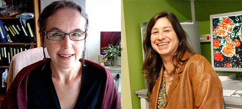
Leslie Griffith & Gina Turrigiano-2017 AAAS Fellows
Leslie Griffith (left) and Gina Turrigiano (right)

Leslie Griffith and Gina Turrigiano have been named American Association for the Advancement of Science (AAAS) Fellows for 2017. This is in recognition of their contributions and scientific leadership in the field of Neuroscience.

Leslie Griffith, Nancy Lurie Marks Professor of Neuroscience and Director of the Volen Center for Complex Systems, studies sleep and memory using Drosophila melanogaster.

Gina Turrigiano is the Joseph Levitan Professor of Vision Science. Her lab studies the mechanisms of homeostatic synaptic plasticity and their effects in developing and functioning cortex.

Vice Provost for Research Edward Hackett is also a 2017 AAAS Fellow in the Section on History and Philosophy in Science.

Griffith, Turrigiano, Hackett and the other Fellows for 2017 will be recognized on Saturday, Feb. 17, 2018 at the 2018 AAAS Annual Meeting in Austin, Texas.

Read more at BrandeisNow.

John Lisman (1944-2017)

Chair of Biology Piali Sengupta wrote:

It is with great sadness that I am writing to let you know that John Lisman passed away last night. He passed away peacefully surrounded by his family. John was an influential and creative scientist and a very good friend to all of us in Biology and Neuroscience. We are glad that we had the opportunity to honor him and hear from him at the Volen Retreat last week. He will be much missed.

John’s talk at the Volen Retreat earlier this month, delivered by video conference, is available here: The critical role of CaMKII in memory storage: 6 key physiological and behavioral tests

The family has asked that in lieu of flowers people consider contributing to the John Lisman Memorial Scholarship

Earlier tweets from past students and colleagues:

https://twitter.com/rennocosta/status/923634257137938434

We also received this longer tribute from Michael Kahana:

I was greatly saddened to hear the news that John Lisman passed away this weekend. I spoke with him just a few weeks ago and was greatly looking forward to his upcoming visit to Penn. Although he told me of his illness, I was hoping to have a little more time with my good colleague and friend. Upon learning of his passing, I wanted to write down a few memories to share with friends and colleagues who knew John well.

I vividly recall when I first met John, at an evening gathering at his home that I attended just prior to joining the faculty at Brandeis (this may have been a precursor to the famous Boston Hippocampus meetings that John helped organize). The meeting was teaming with energy, and John welcomed me warmly, introducing me to other scientists in the room. John had recently become very interested in human memory, and as a newly minted PhD working on memory, John took me under his wings, teaching me about neurophysiology and quizzing me enthusiastically about the psychology of memory, a field that John was keen to master as quickly as possible.

John was a polymath, bursting with creative energy, and capable of seeing connections between diverse fields. Over the subsequent decade at Brandeis, John had an enormous influence on my career and research direction, introducing me to theta and gamma rhythms, and teaching me about countless topics in neurophysiology. On a typical day in the Volen Center, John would rush into my lab eager to share a new discovery or ask me a question about a study of memory that he had just learned about. He had this incredibly-infectious scientific curiosity, and he was always abundantly generous with his time, both with me and my students.

I particularly remember the early days when John was developing the LIJ (Lisman-Idiart-Jensen) model, and trying to learn as much as he could about the Sternberg task and other related phenomena in the field of human memory. Although I frequently challenged John on this front, he kept at it, continuing to refine the model together with Ole Jensen until they were able to answer many of the most serious objections. I just saw that the original paper was cited more than 1,200 times, and several of the follow up papers are well into the many hundreds of citations. This is arguably the most creative neurophysiological model of a cognitive function, and the best example of an attempt to link detailed physiological measurements to behavioral measures of human memory.

We have all lost a great friend, colleague, and mentor, and the field of neuroscience has lost one of its shining stars. I want to share my deepest sympathies with all of you who knew and loved John.

May his family be comforted among the mourners of Zion and Jerusalem.
Mike Kahana

Thomas Reese shared his thoughts:

John, your intellect and spirit lighted more than 30 summers my life at the MBL in Woods Hole.  You were a reference point for neurobiology there, holding court at your favorite table at the Kidd, at the far end of the dock.  A cherished invitation to lunch at exactly 12:00, with interesting synapse people passing though, or to hear a deluge of you new ideas about how a synapse is, or should be, put together.  Occasionally an invitation to dinner outside, behind your house with talk of many things…..joined by the delightful Natashia and other interesting people….discussing well into the night.

If Woods Hole is a little scientific Athens, you were our Socrates, lurking on Milfield. questioning in your disarming, open open way…bringing out the truth.  You were our Dogenes. searching Gardner Road for a man with the honest truth.

John, ,…John..it will seem empty there without you…you
will be very much missed..Tom Reese.     NIH

CaMKII: some basics to remember

The theme of Thursday’s Volen Center for Complex Systems annual retreat will be Breakthroughs in understanding the role of CaMKII in synaptic function and memory and honors the pioneering work of John Lisman. To help bring non-experts up to speed, we asked Neuroscience Ph.D. students Stephen D. Alkins and Johanna G. Flyer-Adams from the Griffith lab at Brandeis for a quick primer on CaMKII.

What’s a protein kinase? 

Protein kinases are enzymes that act by adding phosphate groups to other proteins – a process called phosphorylation. Phosphorylation of a protein usually initiates a cascade of downstream effects such as changes in the protein’s 3D shape,  changes in its interactions with other proteins, changes in its activity and changes in its localization. In causing these types of changes, kinases facilitate some of the most essential cellular and molecular processes required for survival and proper functionality.

Aren’t there lots of protein kinases? What makes CaMKII special? 

Among the roughly 500+ genes in the human genome encoding protein kinases, a kinase known as calcium (Ca2+)/calmodulin-dependent protein kinase II (CaMKII) phosphorylates serine or threonine residues in a broad array of target proteins.  Though found in many different tissues (skeletal muscle, cardiac muscle, spleen, etc.), there is a lot of CaMKII in the brain– about 1% of total forebrain protein and 2% of total hippocampal protein (in rats). Previous research, including pivotal contributions from the Lisman Lab at Brandeis University working in mammalian brain, has identified CaMKII as a cellular and molecular correlate of learning and memory through its multiple roles governing normal neuronal structure, synaptic strength, plasticity, and homeostasis. The Griffith Lab has been instrumental in demonstrating that these roles of the kinase are conserved in invertebrates.

Why do we think CaMKII might play a role in memory?

a) Location!

As previously mentioned, CaMKII accounts for up to 2% of all proteins in memory-important brain regions like the hippocampus. It’s also highly abundant at neuronal synapses, where neurons communicate with each other.

b) Function!

Memory is thought to require a process called long term potentiation (LTP) where two neurons, in response to environmental changes, will change the strength of the synaptic connections by which they communicate with each other—these changes will last even after the environmental input has disappeared. We know that CaMKII is required for LTP. We also know that the increases in neuronal calcium levels that accompany neuronal activation and cause LTP also allow CaMKII to phosphorylate itself. This autophosphorylation of CaMKII changes its kinase activity so that CaMKII can stay active well past the window of neuronal activation, essentially ‘storing’ the memory of previous neuronal activity—much like LTP!

c) Structure!

Ultimately, the issue with ‘molecular memory’ is that all proteins degrade over time, causing one to ask how we can remember things for so long when the original proteins that stored that memory no longer exist. CaMKII is such an exciting candidate for molecular memory because it is mostly found as a dodecameric holoenzyme—this means that CaMKII likes to exist as a big assembly of twelve identical CaMKII subunits. However, each CaMKII subunit retains its kinase activity even when all twelve are assembled. What’s interesting is that the autophosphorylation and activation of one CaMKII subunit (which happens when neurons are activated and intracellular calcium levels rise) actually makes it easier for the other CaMKII subunits in the twelve-unit holoenzyme to become autophosphorylated and activated. This means that maybe when an activated subunit is old and get degraded, another new CaMKII subunit could take its place among the twelve-unit holoenzyme—and become activated just like the old subunit, allowing for the ‘molecular memory’ to last beyond when proteins degrade!

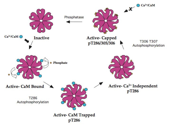
CaMKII phosphorylation and activationCaMKII in more detail…

Calcium binds to the small protein calmodulin and forms (Ca2+/CaM), which acts as a ‘second messenger’ that increases in concentration when neurons are activated. CaMKII relies on calcium/calmodulin (Ca2+/CaM) binding to activate an individual domain containing a regulatory segment.  In conditions of low calcium, elements within the CaMKII regulatory segment will have less affinity for (Ca2+/CaM) binding, keeping CaMKII in an autoinhibited state.  In conditions of high calcium, (Ca2+/CaM) binding initiates phosphorylation at three threonine residue sites, including Thr286 which prevents rebinding of the regulatory segment, thus keeping CaMKII constitutively active even when calcium levels fall.  In this activated state CaMKII can autophosphorylate inactivated intra-kinase domains, and will undergo subunit exchange with neighboring inactivated CaMKII holoenzymes. Furthermore, mutation of CaMKII residues or binding sites in target proteins, such as postsynaptic glutamate (AMPA) receptors, disrupts establishment of long-term potentiation (LTP) in neurons.  Together, CaMKII’s role as molecular switch that bidirectionally, and autonomously regulates activity in neurons has earned it the illustrious title of a “memory molecule.”

What amino-acid manipulations might I hear about?

a) T286A:

Changing a threonine in a phosphorylation site to an alanine prevents phosphorylation at that site. Blocking Thr286 phosphorylation with a T286A mutation prevents CaMKII generation of autonomous activity that disrupts neuronal activity and results in learning deficits.

b) T286D:

Changing a threonine to an aspartate puts a negative charge at the site, often making it act like it’s always phosphorylated. In the case of CaMKII, a T286D mutation renders the kinase constitutively active, which can interrupt normal LTP induction and normal memory storage and acquisition.

To learn more:

Sleep and memory are connected by a pair of neurons in Drosophila

In a recent post on the Fly on the Wall blog, Neuroscience grad student Bethany Christmann talks about recently published research from Leslie Griffith’s lab:

 … [How are sleep and behavior] connected in the brain? Does sleep simply permit memory storage to take place, such that the part of the brain involved in memory just takes advantage of sleep whenever it can? Or are sleep and memory physically connected, and the same mechanism in the brain is involved in both? In a recent study published in eLife, researchers in the Griffith lab may have [uncovered the answer]. They found that a single pair of neurons, known as the DPM neurons, are actively involved in both sleep and memory storage in fruit flies.

Haynes PR, Christmann BL, Griffith LC. A single pair of neurons links sleep to memory consolidation in Drosophila melanogaster. eLife. 2015;4.

Can Self-Referencing Contribute to Memory Errors?

A recent paper in the Journal of Gerontology by Brandeis Ph.D. program alumnus Dr. Nicole Rosa and Professor Angela Gutchess attempts to answer this question. During an interview with ElderBranch, Dr. Nicole Rosa discusses the relationship between self-referencing and false memory. For more information, please read the article on ElderBranch.

To sleep, perchance to learn?

Sleep deprivation is ubiquitous in today’s society, and we have all felt the effects of sleep loss on our ability to function optimally, physically and especially mentally. In particular, it has become clear that the brain requires sleep to efficiently establish many forms of long-term memory. However, it is still unknown what sleep deprivation actually does to the brain to impair its function. In a recently published review in the journal Cellular Signalling, authors Christopher G. Vecsey from Brandeis University and Robbert Havekes and Ted Abel from the University of Pennsylvania have tried to capture the current state of our knowledge about the molecular and cellular effects of sleep deprivation that could explain why sleep loss is so detrimental for memory formation. The review focuses primarily on memories for events and places, which are thought to be formed and stored in the area of the brain called the hippocampus.

A key approach to learn about the nitty-gritty effects of sleep deprivation has been research in rodents. Therefore, the authors begin by summarizing how sleep deprivation studies are carried out in rodents, and how sleep deprivation affects memory and several signaling pathways in the brain. Notably, they review the effects of sleep loss on neurotransmitter systems such as acetylcholine, glutamate, and GABA, all of which could potentially modulate learning and memory. The authors also discuss some of the newest and most exciting studies on the topic of sleep loss, including a handful of experiments in which researchers have been able to reverse the effects of sleep deprivation through pharmacological treatments. For example, the authors describe one of their own studies in which sleep deprivation in mice caused memory deficits and reduced signaling through the cAMP pathway, which is known to be crucial for long-term memory. This molecular effect was likely caused by accelerated breakdown of cAMP by phosphodiesterase 4 (PDE4). When mice were treated with a PDE4 inhibitor during the period of sleep deprivation, memory formation remained unaffected. Rescue of memory defects were also obtained in separate studies in which rodents were treated either with nicotine, caffeine, or CPT, an antagonist of the adenosine A1 receptor. Two related studies also found that the effects of sleep deprivation on memory could be ameliorated by prevention of transmitter release from cells in the brain called glia. This was the first indication that brain cells other than neurons are impacted by sleep deprivation and that they contribute to the effects of sleep loss on the ability to remember new information.

As the authors mention, goals for studies in the immediate future will be to identify additional ways that sleep deprivation affects the brain, determine why sleep deprivation targets these molecules, and discover how these targets interact with each other to impair the normal function of the brain. Finally, hopefully our growing knowledge can be used to develop treatments for the cognitive deficits produced by sleep loss in people, especially those who have impaired sleep due to a medical condition, such as insomnia, chronic pain, sleep apnea, or one of the many neurodegenerative or psychiatric disorders associated with disturbed sleep patterns.

Christopher G. Vecsey is a postdoctoral fellow in the Griffith Lab at Brandeis, where he continues to work on interactions between sleep and learning. Chris is supported by a postdoctoral fellowship from the National Institute of Mental Health.

What we can learn about aging from worms

Coleen Murphy from the Dept of Molecular Biology at Princeton will tell us about “Slowing the Ticking Clock: What we can learn about aging and memory from C. elegans at the first Ruth Ann and Nathan Perlmutter Science Forum on Wednesday, March 9 at 4:00 pm in Gerstenzang 121. The focus of her research is on understanding the genes that regulate longevity, using C. elegans as a model system. Coleen performed her Ph.D. thesis research with Jim Spudich at Stanford where she studied myosin motors and then went on to  a post doctoral fellowship with Cynthia Kenyon at UCSF where she began studying aging. Since starting her own lab at Princeton, Coleen has been the recipient of numerous prestigious awards including a Pew Scholar Award, a Keck Distinguished Young Scholar Award, and an NIH Director’s Innovator Award. Her lab’s most recent work showed that TGF-β and insulin signaling regulates reproductive aging. In addition, her lab has also recently been looking into the connection between longevity mutants and memory in C. elegans

About the Forum: Ruth Ann Perlmutter has been a longtime friend of Brandeis University. In 1969, Nathan Perlmutter became vice president of development at Brandeis during the presidency of Morris Abrams. Perlmutter left Brandeis to become the National Director of the Anti Defamation League. Together the Perlmutters were leaders in the interfaith movement and civil rights debates for which activities Nathan received the Presidential Medal of Freedom shortly before his death in 1987. Mrs. Perlmutter earned her B.A. from the University of Denver and her masters degree in sociology from Wayne State University in Detroit. She is a sculptor and painter in her own right and currently lives in Prescott, Arizona.

Wingfield Receives 2010 Baltes Distinguished Research Achievement Award

Update; BrandeisNOW has a in-depth profile on Prof. Wingfield.

Professor Arthur Wingfield is the 2010 recipient of the Baltes Distinguished Research Achievement Award. The $5000 award, given annually by the Margaret M. and Paul B. Baltes Foundation and Division 20 (Adult Development and Aging) of the American Psychological Association (APA), recognizes outstanding contributions to our understanding of adult development and aging. As part of the award, Wingfield will deliver a keynote address at the next annual meeting of the APA.

The number of adults age 65 or older in the US is expected to grow from 35 million in the year 2000, to 70.3 million in 2030.  Among this group, hearing loss is the third most prevalent chronic medical condition, exceeded only by arthritis and hypertension.  The hearing loss associated with adult aging, or presbycusis (literally, “old hearing”) presents a more complex picture than many realize. Whether the loss is mild or more severe, the source is a thinning of hair cells located in the cochlea, a spiral-shaped structure about only the size of the nail on your little finger. There are also “higher level” effects that include the pathways from the cochea to the brain, and age-related changes in the auditory receiving areas of the brain itself. These biological changes result in the older listener expending attentional effort that is not only tiring, but can draw on resources that would ordinarily be available for encoding what has been heard in memory.

This recent award recognizes Wingfield and his Brandeis colleagues’ contributions to understanding this complex interaction between sensory and cognitive changes in adult aging.  Arthur Wingfield is the Nancy Lurie Marks Professor of Neuroscience and director of the Volen National Center for Complex Systems at Brandeis.  His work has also been recognized by the American Speech, Language and Hearing Association, and two successive MERIT Awards from the NIH’s National Institute on Aging.


Brandeis hosts International Workshop on Learning and Memory

25 internationally recognized scientists gathered at Brandeis University to discuss recent progress in understanding the neural mechanisms that promote learning. The workshop was sponsored by the Science of Learning Division of the National Science Foundation in a grant to Brandeis University Professor John Lisman, the Zalman Abraham Kekst Chair in Neuroscience. Lisman and Dr. Emrah Duzel, a neurologist from University College London, were the co-organizers of the workshop. Among the leading scientists attending were Mortimer Mishkin, Chief of the Cognitive Section on Neuroscience at NIMH, and the Nobel Prize recipient, Susumu Tonegawa.

The question of how the brain changes during learning has long fascinated scientists. In 1949 the Canadian psychologist, Donald Hebb, proposed that learning new associations involves changes in the strength of synapses. Subsequent work in many laboratories established that synapses do change as we learn and that the process rather closely follows the specific rule that Hebb had postulated. Recent work, however, has revealed a limitation of Hebb’s rule; the forming of associations depends on the novelty of incoming information and on the motivation to learn, factors that Hebb’s rule cannot account for. The purpose of the workshop was to see how Hebb’s rule could be revised to take into consideration the new findings […]

25 internationally recognized scientists gathered at Brandeis University from October 3-5, 2010, to discuss recent progress in understanding the neural mechanisms that promote learning. The workshop was sponsored by the Science of Learning Division of the National Science Foundation in a grant to Brandeis University Professor John Lisman, the Zalman Abraham Kekst Chair in Neuroscience. Lisman and Dr. Emrah Duzel, a neurologist from University College London, were the co-organizers of the workshop. Among the leading scientists attending were Mortimer Mishkin, Chief of the Cognitive Section on Neuroscience at NIMH, and the Nobel Prize recipient, Susumu Tonegawa.

The question of how the brain changes during learning has long fascinated scientists. In 1949 the Canadian psychologist, Donald Hebb, proposed that learning new associations involves changes in the strength of synapses. Subsequent work in many laboratories established that synapses do change as we learn and that the process rather closely follows the specific rule that Hebb had postulated.  Recent work, however, has revealed a limitation of Hebb’s rule; the forming of associations depends on the novelty of incoming information and on the motivation to learn, factors that Hebb’s rule cannot account for. The purpose of the workshop was to see how Hebb’s rule could be revised to take into consideration the new findings.
[…]