Simulations Say Viral Genome Lengths are Optimal for Capsid Assembly

Viruses are infectious agents made up of proteins and a genome made of DNA or RNA. Upon infecting a host cell, viruses hijack the cell’s gene expression machinery and force it to produce copies of the viral genome and proteins, which then assemble into new viruses that can eventually infect other host cells. Because assembly is an essential step in the viral life cycle, understanding how this process occurs could significantly advance the fight against viral diseases.

In many viral families, a protein shell called a capsid forms around the viral genome during the assembly process. Capsids can also assemble around nucleic acids in solution, indicating that a host cell is not required for their formation. Since capsid proteins are positively charged, and nucleic acids are negatively charged, electrostatic interactions between the two are thought to be important in capsid assembly. Current questions of interest are how structural features of the viral genome affect assembly, and why the negative charge on viral genomes is actually far greater than the positive charge on capsids. These questions are difficult to address experimentally because most of the intermediates that form during virus assembly are too short-lived to be imaged.

hagan-capsid-sim
Snapshots from a computer simulation in which model capsid subunits (blue) assemble around a linear, negatively charged polymer (red). Positive charges on the capsid proteins are shown in yellow.

In a new paper in eLife, Brandeis postdoc Jason Perlmutter, Physics grad student Cong Qiao, and Associate Professor Michael Hagan have used state of the art computational methods and advances in graphical processing units (on our High Performance Computing cluster) to produce the most realistic model of capsid assembly to date. They showed that the stability of the complex formed between the nucleic acid and the capsid depends on the length of the viral genome. Yield was highest for genomes within a certain range of lengths, and capsids that assembled around longer or shorter genomes tended to be malformed.

Perlmutter et al. also explored how structural features of the virus — including base-pairing between viral nucleic acids, and the size and charge of the capsid — determine the optimal length of the viral genome. When they included structural data from real viruses in their simulations and predicted the optimal lengths for the viral genome, the results were very similar to those seen in existing viruses. This indicates that the structure of the viral genome has been optimized to promote packaging into capsids. Understanding this relationship between structure and packaging will make it easier to develop antiviral agents that thwart or misdirect virus assembly, and could aid the redesign of viruses for use in gene therapy and drug delivery.

Perlmutter JD, Qiao C, Hagan MF. Viral genome structures are optimal for capsid assembly. eLife 2013;2:e00632

Prolonging assembly through dissociation

Microtubules are semiflexible polymers that serve as structural components inside the eukaryotic cell and are involved in many cellular processes such as mitosis, cytokinesis, and vesicular transport. In order to perform these functions, microtubules continually rearrange through a process known as dynamic instability, in which they switch from a phase of slow elongation to rapid shortening (catastrophe), and from rapid shortening to growth (rescue). The basic self-assembly mechanism underlying this process, assembly mediated by nucleotide phosphate activity, is omnipresent in biological systems.  A recent paper, Prolonging assembly through dissociation: A self-assembly paradigm in microtubules ,  published in the May 3 issue of Physical Review E,  presents a new paradigm for such self-assembly in which increasing depolymerization rate can enhance assembly.  Such a scenario can occur only out of equilibrium. Brandeis Physics postdoc Sumedha, working with Chakraborty and Hagan, carried out theoretical analysis of a stochastic hydrolysis model to demonstrate the effect and predict features of growth fluctuations, which should be measurable in experiments that probe microtubule dynamics at the nanoscale.

Model for microtuble dynamics. All activity is assumed to occur at the right end of the microtubule (denoted as ">")

The essential features of the model that leads to the counterintuitive result of depolymerization helping assembly are (a) stochastic hydrolysis that allows GTP to transform into GDP  in any part of the microtubule, and (b) a much higher rate of GTP attachment if the end of the microtubule has a GTP-bound tubulin dimer, compared to a GDP-bound tubulin dimer.    Process (a) leads to islands of GTP-bound tubulins to be buried deep in the microtubule.   Depolymerization from the end reveals these islands and enhances assembly because of the biased attachment rate (b).  The simplicity of the model lent itself to analytical results for various aspects of the growth statistics in particular parameter regimes.   Simulations of the model supported these analytical results, and extended them to regimes where it was not possible to solve the model analytically.  The statistics of the growth fluctuations in this stochastic hydrolysis model are very different from “cap models” which do not have GTP remnants buried inside a growing microtubule.   Testing the predictions in experiments could, therefore, lead to a better understanding of the processes underlying dynamical instability in-vivo and in-vitro.   An interesting question to explore is whether the bias in the attachment rates is different under different conditions of microtubule growth.

Molecular mechanisms of noisy transcription

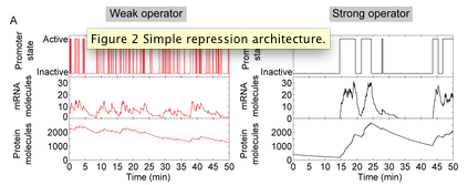
In a recently published paper “Effect of promoter architecture on the cell-to-cell variability in gene expression” in PLoS Computational Biology, Alvaro Sanchez and co-workers investigated how the architecture of a model promoter region (characterized by number of transcription factor binding sites, the binding affinity and spacing on the DNA) affects the way in which individual cells respond to environmental stimuli. In particular, they examine, using stochastic chemical kinetics, how the intrinsic randomness in the binding and unbinding of transcription factors to their binding sites generates cell-to-cell differences in transcript and protein levels within a population. The analysis uses a combination of computational modeling and analytical mathematical methods. Sanchez, a recent Ph.D. graduate in Biophysics and Structural Biology performed this work with Jané Kondev (Physics), and in collaboration with Rob Phillips, Hernan Garcia and Daniel Jones (Caltech).

While previous population-average models explained well how promoter architecture affects the average response of a population of cells to changes in the concentration of transcription factors, the question of how the response of individual cells is determined by promoter region sequence remains generally unsolved and limited to simplified coarse-grain models. By way of an example, the authors of this study investigated the effect of cooperative binding between transcription factors in the level of variability in the transcriptional response to increasing concentrations of those factors. It is well known that cooperativity in gene regulation increases the sensitivity of the response of the promoter to changes in the intracellular concentration of transcription factors, leading to a switch-like response. By examining this architecture, Sanchez and co-workers found that cooperativity is also a source of large intrinsic cell-to-cell variability in gene expression: larger sensitivity comes accompanied with larger variability (even if all cells contain the exact same amount of repressor).

This investigation continues a collaboration between theorists at Brandeis and experimentalists at Caltech, which aims to connect the biochemical, molecular understanding of transcriptional regulation coming from in vitro biochemical experiments (which are also being done in the Gelles lab at Brandeis) with the phenotypic behavior of individual cells as determined by gene expression measurements in single live cells. Many of the predictions of this computational study are currently being tested in Rob Phillips’ lab.


Quantitative Biology Bootcamp 2011

The 5th Annual Quantitative Biology Bootcamp will be held on January 16 & 17, 2011.  Paul Miller will preside over the 2nd annual QB Computational Challenge:  When space trumps time: modeling dynamic spatial patterns with Matlab. This year’s panel discussion topic is “Writing interdisciplinary papers. What to do. What not to do.”  We’re delighted to announce the HHMI Interfaces Scholar award went to Adelajda Zorba (Kern lab).  Adelajda was selected from among several exceptional submissions this year.  The topic is HIV-1 assembly.

Members of the Brandeis community are invited to attend. If you are interested, please contact Trisha Murray no later than Jan. 4, 2011.

HPC cluster hits milestone

Our high-performance computing cluster passed the 1000 core mark this month, thanks to computer purchases for the computational biophysics and computational neuroscience groups and infrastructure support from Library and Technology Services. I’m looking forward to another great year working with you all.

BLAST (1): New versions

NCBI has released new versions of their web and standalone BLAST+ programs. This appears to me significant mostly if you BLAST lots of sequences. I used to have a computer with the standalone programs installed, but no one was using it. If someone needs BLAST+ installed on a local computer, let me know.